
Differentiating the mild and severe ends of the same infection — symptoms, workup, treatment, and prognosis
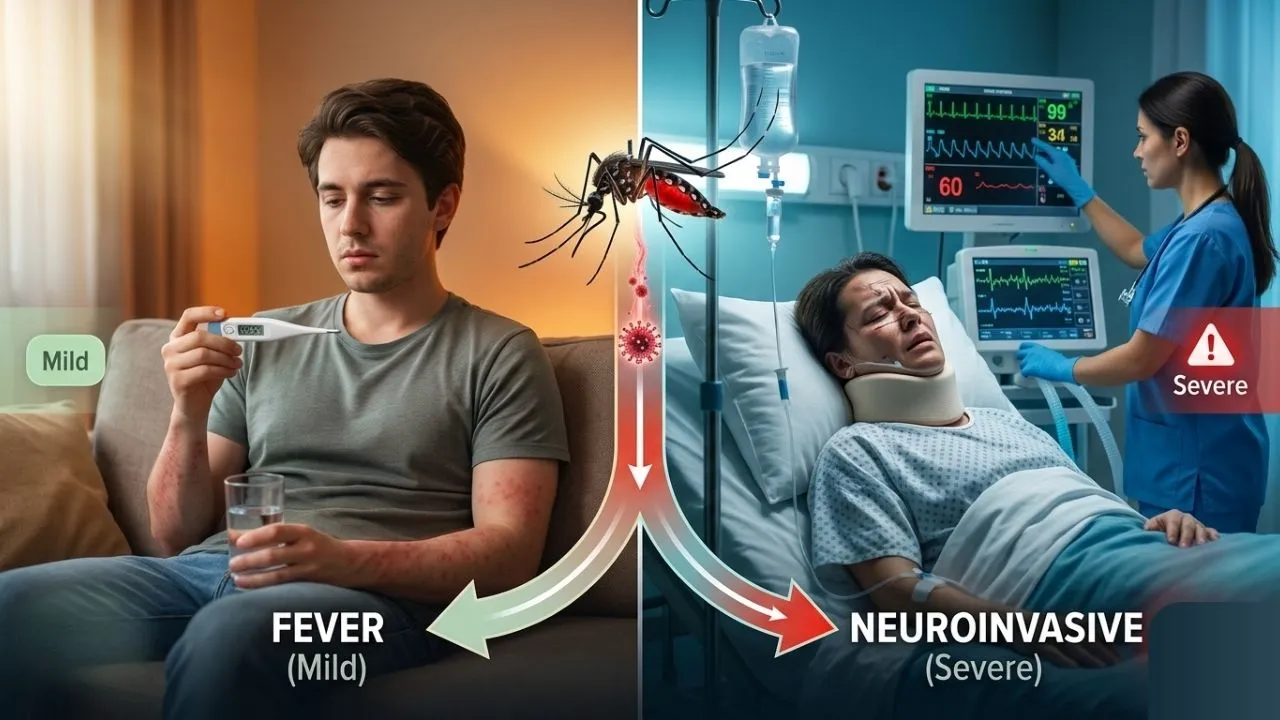
Two patients walk into an ED in the same August week. Same mosquito season, probably the same neighborhood, maybe even the same species of Culex behind it. One has a fever, a pounding headache, and body aches that resolve in five days without ever seeing a doctor. The other is confused, running a high fever, and can’t lift her right leg off the bed.
Same virus. Wildly different disease.
That gap — between West Nile fever and West Nile virus neuroinvasive disease — is one of the more clinically important distinctions in arboviral medicine, and it doesn’t get explained clearly enough. Most patient-facing content flattens it into “mild or severe” and moves on. That’s not really adequate if you’re a clinician trying to triage a summer febrile illness, or a patient trying to understand why your cousin’s case looked nothing like yours.
This is a full clinical comparison — pathophysiology, symptom differentiation, diagnostic workup, treatment, and prognosis — pulled from CDC clinical guidance, StatPearls, UpToDate, and peer-reviewed case series. Let’s get into it.
West Nile Fever vs. West Nile Virus Neuroinvasive Disease: The Core Distinction
What is the Difference Between West Nile Fever and West Nile Neuroinvasive Disease?
Both conditions come from the same infection. What differs is whether the virus stays confined to a systemic, self-limited illness, or crosses into the central nervous system.
- West Nile fever (WNF) — the non-neuroinvasive form. A systemic, flu-like illness. Self-limited. No CNS involvement.
- West Nile virus neuroinvasive disease (WNND) — the virus crosses the blood-brain barrier, producing meningitis, encephalitis, acute flaccid paralysis, or some combination
The split in who gets which is fairly well established at this point. Roughly 80% of infections are asymptomatic. Of the remainder, about 20 to 25% develop West Nile fever. Only 1 in 150 to 250 infected people — under 1% of total infections — progress to neuroinvasive disease. It’s rare. It’s just not rare enough to ignore.
Pathophysiology: Why Some Infections Stay Systemic and Others Don’t
Understanding why one patient gets a headache and another gets encephalitis starts with what the virus is actually doing at the cellular level.
West Nile virus is a Flaviviridae family arbovirus, transmitted through the bite of an infected Culex mosquito. After inoculation, the virus initially replicates in skin dendritic cells and regional lymph nodes, then disseminates through the bloodstream — this is the viremic phase that produces the systemic symptoms of West Nile fever.
For most patients, the immune system contains the infection at this stage. The virus is cleared, symptoms resolve, and it never gets anywhere near the brain.
In a small subset, though, the virus crosses the blood-brain barrier. The exact mechanism isn’t fully settled, but the leading theories include cytokine-mediated increases in vascular permeability, infected macrophages trafficking the virus across the barrier directly, and retrograde axonal transport through olfactory or peripheral neurons. Once inside the CNS, persistent viral replication can occur, which is part of why neurologic sequelae can linger well past the acute illness.
The paralysis seen in acute flaccid myelitis has a specific mechanism worth calling out: it results from destruction of the anterior horn cells of the spinal cord — the same cells targeted in poliomyelitis, which is exactly why WNV-associated paralysis can look so similar to polio on exam.
Risk Factors for Progression to Neuroinvasive Disease
- Age — by far the strongest predictor; risk climbs sharply after 60, especially for encephalitis.
- Immunosuppression — organ transplant recipients and immunocompromised patients show significantly higher rates of ICU admission, mechanical ventilation, and mortality.
- Diabetes and hypertension — both associated with increased neuroinvasive risk in cohort data.
- Higher initial viral load — some evidence links viral burden to fever severity, and possibly to progression risk.
None of these are hard rules. Healthy, younger patients do occasionally develop neuroinvasive disease. But if you’re risk-stratifying a febrile patient in mosquito season, age and immune status should be the first two boxes you check.
Symptom Differentiation: West Nile Fever
This is the illness most symptomatic patients experience, and it’s genuinely just uncomfortable rather than dangerous.
Common Presenting Symptoms
- Sudden-onset fever
- Headache — typically diffuse, not the severe, distinct quality seen in meningitis
- Myalgia and arthralgia (muscle and joint pain)
- Fatigue, sometimes profound
- Gastrointestinal symptoms — nausea, vomiting, or diarrhea
- Maculopapular rash — seen in roughly half of symptomatic patients, usually on the trunk and extremities
- Lymphadenopathy, less commonly
It’s worth noting: fever isn’t universal even here. About 4% of WNV disease cases present without fever at all, which is part of why the CDC revised its surveillance case definition back in 2013 to stop requiring fever as a strict criterion for neuroinvasive disease.
Clinically, this is a self-limited illness, per StatPearls guidance, with most symptoms resolving within about 10 days. Fatigue tends to outlast everything else, sometimes by weeks.
Symptom Differentiation: West Nile Virus Neuroinvasive Disease
This is where the clinical picture changes meaningfully — not just in intensity, but in kind.
Meningitis Presentation
- Fever, often high
- Severe headache, distinct from a typical tension-type or systemic headache
- Neck stiffness (nuchal rigidity)
- Photophobia
- Generally similar clinical picture to other viral meningitides — patients are uncomfortable, but mental status stays largely intact
Meningitis is seen more often in children, and generally carries the best prognosis of the three neuroinvasive presentations.
Encephalitis Presentation
- Altered mental status sustained beyond 24 hours — this is part of the formal case definition
- Confusion, disorientation, lethargy
- Seizures
- Focal neurologic findings
- In one NYC case series, altered mental status was present in 72.2% of neuroinvasive cases, and lethargy in 63.9%
Encephalitis skews toward an older population and carries the more serious prognosis of the neuroinvasive presentations. It’s also, worth noting, the presentation most often actually meningoencephalitis on closer inspection — CSF findings frequently mirror meningitis even when altered mental status dominates the clinical picture.
Acute Flaccid Paralysis (Acute Flaccid Myelitis)
- Asymmetric, painless limb weakness — the hallmark feature
- Hyporeflexic or areflexic weakness on exam, generally with intact sensation
- Can occur in isolation or alongside encephalitis or meningitis
- Sometimes preceded by localized back pain before weakness sets in
- Bowel, bladder dysfunction, and respiratory compromise requiring ventilatory support in severe cases
- Occasionally mimics Guillain-Barré syndrome, with peripheral demyelination rather than the classic anterior horn cell pattern
This presentation is genuinely tricky diagnostically. It can appear weeks after the initial illness, and in some documented cases, weakness has relapsed after a period of apparent improvement — an atypical temporal pattern that catches clinicians off guard if West Nile isn’t already on the differential.
Common Symptoms Across All Neuroinvasive Presentations
A few symptoms show up broadly across meningitis, encephalitis, and AFP, regardless of which dominates the picture:
- Fever, headache, anorexia
- Nausea or vomiting
- Gait disturbance and walking instability
- Memory impairment, difficulty concentrating
- Tremor, and less commonly diplopia or nystagmus
- Depression, during both the acute phase and recovery
WNV & WNND | Diagnostic Workup: How Clinicians Tell Them Apart
Diagnosis, honestly, is the part where this disease trips people up the most — including experienced clinicians. One Arizona outbreak review found that only 40% of patients with clinically compatible meningitis or encephalitis were even tested for WNV. It requires a high index of suspicion, especially outside peak mosquito season or in regions where WNV isn’t top-of-mind.
Serologic Testing
- IgM antibody-capture ELISA (MAC-ELISA) on serum is the standard first-line test
- IgM typically becomes detectable within 8 days of symptom onset — testing too early can produce a false negative, and a repeat test 10 days later is standard practice if suspicion remains high
- IgM can persist in blood for up to 500 days, which complicates using a single sample to confirm acute infection — paired acute and convalescent samples are often needed
- Cross-reactivity with other flaviviruses (dengue, Zika, St. Louis encephalitis) can cause false positives; the plaque reduction neutralization test (PRNT) helps resolve ambiguous results
Cerebrospinal Fluid (CSF) Analysis — Required for Suspected Neuroinvasive Disease
Any patient with neurologic symptoms in the appropriate clinical context should get a lumbar puncture. Typical findings:
- Elevated protein, often in the 100–1000 mg/dL range
- Pleocytosis with lymphocytic predominance — though early in the course, a neutrophilic predominance is possible and can be a diagnostic pitfall
- Generally normal glucose
- CSF IgM testing via ELISA is more specific for CNS infection than serum alone, since IgM antibody doesn’t cross the blood-brain barrier — a positive CSF IgM is stronger evidence of true intrathecal infection
- PCR can detect WNV RNA directly, useful early in infection before antibodies form, but sensitivity is limited — roughly 55% in CSF and only about 10% in blood
A typical CSF findings do happen — cases with elevated protein but minimal pleocytosis (albuminocytologic dissociation), or unusual neutrophilic predominance, have been documented and can mimic other conditions like Guillain-Barré syndrome. Clinical correlation always matters more than any single lab value in isolation.
Neuroimaging
- CT of the brain is typically normal in acute WNV disease and adds little diagnostic value on its own
- MRI is more useful — abnormal findings appear in roughly one-third of neuroinvasive cases
- Classic MRI findings include T2 hyperintensities in the bilateral thalami, brainstem, and deep white matter
- Leptomeningeal and periventricular enhancement can be seen with contrast
- In suspected AFP or myelitis, MRI of the spine may show enhancement of the ventral nerve roots or cauda equina — sometimes the only imaging abnormality present, even when brain MRI is unremarkable
West Nile Fever vs. Neuroinvasive Disease: Side-by-Side Comparison
| Feature | West Nile Fever | Neuroinvasive Disease |
|---|---|---|
| Approx. frequency | ~20–25% of infections | <1% (1 in 150–250) |
| CNS involvement | None | Yes — meningitis, encephalitis, and/or AFP |
| Mental status | Normal | Often altered (especially in encephalitis) |
| Typical duration | Up to 10 days; fatigue may persist longer | Weeks to months; hospitalization common |
| Case fatality | Essentially none | ~9–10%, highest in encephalitis and AFP over age 70 |
| LP/CSF required | Not routinely | Yes, for diagnosis and to rule out mimics |
| Highest-risk group | Women, higher viral load (fever risk) | Age 60+, immunosuppressed patients |
West Nile Virus: Treatment Approaches
Here’s the part that surprises a lot of patients: there’s no antiviral drug specific to West Nile virus, for either presentation.
1. Management of West Nile Fever
- Supportive care only — rest, hydration, and over-the-counter analgesics/antipyretics
- Outpatient management in the vast majority of cases
- No indication for antivirals or antibiotics — this is a self-limited viral illness
2. Management of Neuroinvasive Disease
- Hospitalization, often with ICU-level care for encephalitis or AFP with respiratory involvement.
- Supportive management remains the mainstay — IV fluids, seizure control, pain management, and close neurologic monitoring.
- Mechanical ventilation may be required in AFP cases with respiratory muscle involvement.
- Various specific therapeutic agents (interferon, IVIG, corticosteroids) have been trialed and described in case reports, but evidence for efficacy remains limited across the board.
- Immunosuppressed patients require closer monitoring given documented higher rates of severe outcomes.
It’s a genuinely frustrating gap in the treatment landscape. Decades after WNV’s 1999 emergence in New York, management is still built almost entirely around supportive care and close observation rather than targeted antiviral therapy.
West Nile Fever vs. West Nile Virus Neuroinvasive Disease: Prognosis and Long-Term Outcomes
1. West Nile Fever Prognosis
Excellent, across essentially every data source. Most patients recover fully, though fatigue and weakness can persist for weeks to months beyond the acute illness — a detail worth setting expectations around, since it surprises a fair number of patients who assume they should feel back to normal within days.
2. Neuroinvasive Disease Prognosis
Meaningfully more guarded. Case fatality for WNND runs approximately 9 to 10%, and mortality risk climbs further in patients with encephalitis or AFP, particularly those over 70.
- Meningitis generally carries the best prognosis of the three neuroinvasive presentations, with most patients recovering fully
- Encephalitis carries a more serious course, with higher rates of lasting cognitive and functional impairment
- AFP has the most unpredictable recovery arc — some patients regain substantial function through rehabilitation, others are left with permanent weakness
- Long-term follow-up data shows a meaningful proportion of patients — roughly 40% in one 8-year cohort study — report ongoing symptoms years after infection, with encephalitis and age over 50 as the strongest predictors of prolonged recovery
Recovery, even for neuroinvasive patients, is not universally bleak. Interval improvement on repeat imaging and resolution of symptoms at follow-up is well documented, including cases initially presenting with striking MRI abnormalities that improved substantially over months.
Clinical Takeaways: When to Suspect Neuroinvasive Disease
For clinicians managing a febrile patient during mosquito season, a few practical flags matter more than others:
- Any sustained alteration in mental status beyond 24 hours warrants a lumbar puncture, not a wait-and-see approach
- Asymmetric limb weakness, especially painless and with intact sensation, should raise suspicion for WNV-associated AFP, particularly in an epidemiologically consistent setting
- Don’t rely on fever presence or absence alone — a meaningful minority of both fever and neuroinvasive cases present afebrile
- Age over 60 and immunosuppression should lower your threshold for CSF testing in any patient with even mild neurologic complaints
- A single negative IgM early in the illness doesn’t rule out infection — repeat testing at 8–10 days is standard when suspicion remains
The broader point, and it’s one worth sitting with: West Nile fever and West Nile virus neuroinvasive disease are technically the same infection, but clinically, they’re almost different diseases. One resolves at home with fluids and rest. The other can mean a spinal tap, an ICU bed, and a recovery measured in months rather than days. Knowing which one you’re looking at — early, and correctly — is most of what good management here actually comes down to.
Editorial Team & Contributors

Raashid Ansari, Founder, Content Researcher & Author - MosquiTalk
Not an entomologist — just a genuinely curious writer who started researching mosquitoes and couldn't stop. What began as casual reading about repellents and bite prevention gradually turned into a deep ongoing dive into vector biology, disease epidemiology, animal health impacts, and the real science behind mosquito control. Everything published here is carefully edited, and written with one purpose: giving readers accurate, accessible information they can actually trust and use to protect themselves, their families, and their pets, birds and cattle.
Active across social platforms, regularly publishes, and genuinely invested in spreading mosquito awareness where it matters most. Because informed readers make better decisions — and better decisions save lives.